An Interview with Dr. Dennis DePace
By Philip Yates
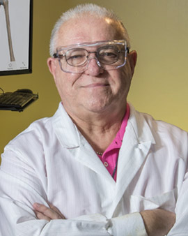
Dr. Dennis DePace has been with Drexel University College of Medicine, formerly Hahnemann Medical College, for more than 40 years as an anatomy professor. In this conversation, he shares his appreciation for human anatomy and his passion for educating the future generation of scientists and physicians in the long tradition of experiential learning.
What brought you here?
In 1974, I completed my PhD at the University of Buffalo, and I attended the American Association of Anatomists meeting. There I met the chairman of anatomy at Hahnemann, who was rebuilding the department of anatomy and was looking for a neuroscientist. My research had been in neuroscience, so he hired me to be the course director of the neuroanatomy course. In subsequent years, I also had experience in gross anatomy. I moved into that and eventually gave up neuro in favor of gross, which is something I like more because there are more organs than the brain, but don't tell the neurobiology group I said that.
How has the department changed over the years?
It changed when Dr. Fischer became chair because he is very supportive of the educational mission in addition to the research. In my previous experience with other iterations of the department, that was not always the case, because the research brings in the money that supports the activities of the department. So it is really great for me now to be a part of a department that is so well funded, has research that is top-notch, and yet also has support for the educational mission.
How has your role in the department changed over the years?
My role hasn't changed that much, but teaching has definitely changed from when I started. When I began my career there was no technology. There were no computers, no internet. All of these things have evolved during my career. It's fun to take advantage of those resources now and incorporate new technologies into my teaching. Dr. Schidlow, as dean of the medical school, has been very supportive; our gross lab was equipped with new computers about four years ago.
What is your favorite thing about working here?
My number one favorite thing are my colleagues because we have a very good working relationship. The faculty and staff are excellent people who interact very well in a very positive way that helps us to be as good as we can. And of course, I love teaching. We have a very diverse student body that makes it interesting to meet students from different backgrounds and cultures. Those are two things that I value the most.
What is the process for a cadaver to come to our lab?
The cadavers come to us from a humanities gift registry that is a central organization in the Commonwealth for handling body donations, so all of our cadavers are willed bodies. When a person who has made a donation passes away, the humanities gift is notified by the family, and they send one of the local undertakers to take the body to one of the medical schools in a rotation, although there are people who specifically will their body to this school. Once the body comes here, we have an embalmer who embalms the body, after which the body is put in cold storage. We usually keep the cadavers in cold storage for six months before we use them, in order to allow more time for the embalming fluid to permeate and for the tissues to be well fixed.
Have any of the cadavers surprised you with something unique or unusual?
I have seen a lot of interesting findings. One that stands out is that one year the students were dissecting the pelvis and something caught my eye. In the bladder were five rocks that were originally bladder stones. They had accumulated over the years to be quite large. This person went around life with these rocks in his bladder. I investigated about it and learned that in the 1800s people were trained to insert instruments into the urethra to crush bladder stones to remove them. Patients were instructed to sit back with the feet in stirrups in what is commonly called the lithotomy position, which is used for gynecological examination today.
Another incident goes back many years, when we had a cadaver of a 95-year-old man at Hahnemann. When we opened his chest and abdomen, we discovered that he had situs inversus totalis with complete reversal of his organs from left to right. It can happen in varying degrees, but this was a total reversal. We had no medical history and didn't know whether he was even aware of it.
What are you working on now?
My current avocation is re-working the anatomy images that were published in the 1916 edition of Gray's Anatomy. They are in the public domain, so we can use them in our lectures without worrying about copyright. They are all black and white, so I am using the free image program Gimp, which is similar to Adobe Photoshop, to colorize and label the images. I uploaded a few of them to Wikimedia Commons so other people can access them. It's fun to see how you can display the illustrations to make certain things pop out, because sometimes it's hard to find good illustrations.
What do you do when you're not teaching?
For down time, I love to cook. I live in a great part of the city close to the 9th street Italian market and the Reading Terminal Market, so there's a ready abundance of ingredients. I have a large collection of cookbooks and I like to try new dishes. I also enjoy photography and gardening. We have a back garden patio in Center City where we do a lot of gardening. I record some of my lectures in my office at home and sometimes I leave my deck door open so you can hear birds chirping in the lectures. Students tell me they like that.
Do you have any advice for young scientists and students?
The anatomy teachers nationally are getting older, and there isn't a whole lot of replacements coming along. I would say if you have the ability to do it, learning anatomy is a good idea as a backup for a research career. Medical schools will continue to teach anatomy, whether it is done electronically or with cadavers. We still believe cadaver dissection is the best way to do it because there is a whole lot that comes from the experience besides learning the anatomy. There is the professionalism element. It is one of the first opportunities for medical students to work as a team on a long-term project. It gives an appreciation of human variation, as not every case you see in the clinic will be identical. You start to appreciate the nuances of different people. It's a unique experience, and most physicians view it as a rite of passage into the profession.