First Place
The Space Base by Alberto Brambila Solórzano, Colima´s University, Mexico.
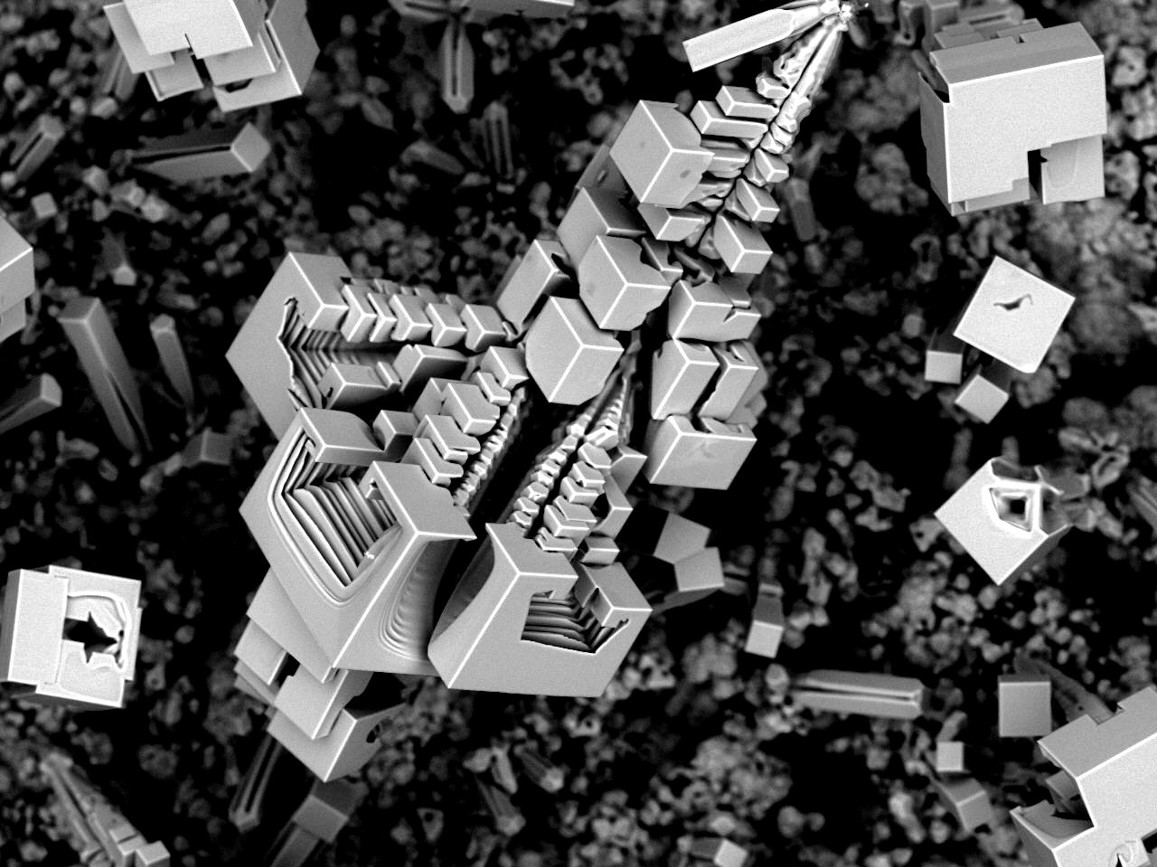
The Spatial Base of NaF is an image obtained by SEM, was synthesized through the CVD method, this microstructure formed by the coupling of the cubes in space, this base seems to be floating in the middle of nowhere waiting for some traveler. Image width is 0.5 mm.
Second Place
Evolution of peptide nanostructures by Charalampos Pappas, Advanced Science Research Center, City University of New York, USA.
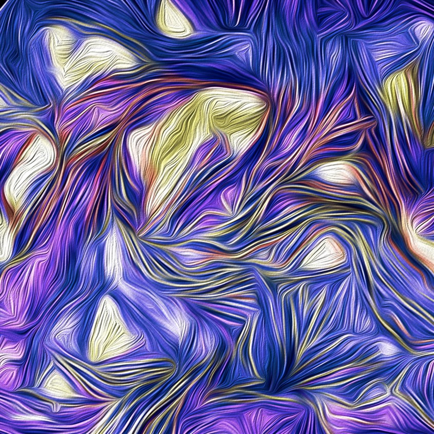
This TEM image represents an example of a supramolecular peptide nanostructure that was discovered using a dynamic peptide library approach, where peptide sequences are dynamically exchanged, giving rise to a competition of sequences and resulting in the spontaneous selection and formation of stable self-assembling nanostructures. Image width is 0.0002mm.
Third Place (tie three ways)
Magnetostatic Spawn by Aleks Labuda, Asylum Research, Santa Barbara, USA.
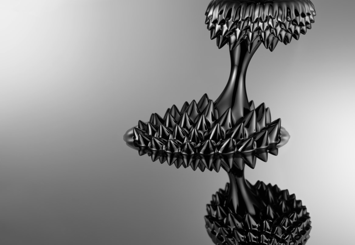
Ferrofluids are colloidal liquids made of nanoscale ferromagnetic particles suspended in a carrier fluid. Surfactants in the fluid prevent the agglomeration of the nanoparticles because their strong Van der Waals forces exceed the particles’ weak magnetic attraction. Reducing the size of the magnetite (or hematite) nanoparticles to below 10 nm is key in preventing their precipitation from the carrier fluid. At such length scales, brownian motion ensures their indefinite suspension under normal conditions. This photograph demonstrates the magnetic properties of this liquid as it shape shifts under the magnetic field setup by two carefully positioned rare-earth disc magnets. One magnet lies below a silicon wafer, while the other hangs from above. The protruding cones are a result of the compromise between the minimization of magnetic energy versus the minimization of surface free energy.
Vanadium Oxide Leaves by Mallory Clites, Drexel University, USA.
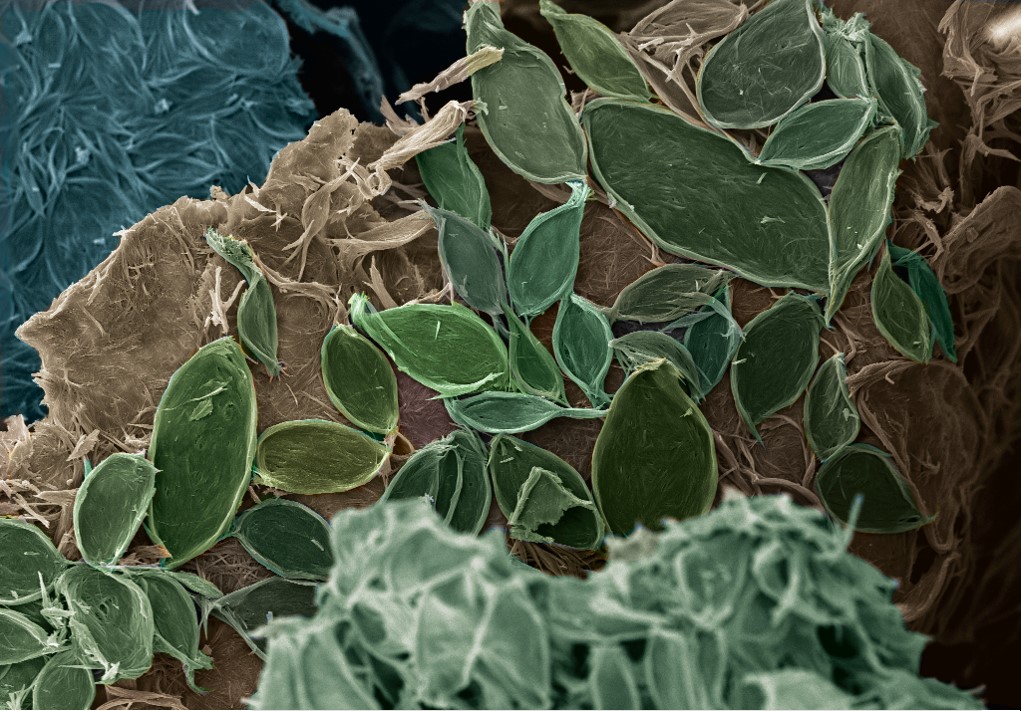
Bilayered vanadium oxide has a flexible layered crystal structure that allows for the intercalation of various ions and molecules. In this image, a large, positively-charged organic molecule, cetyltrimethyl ammonium, has been inserted into the space between vanadium oxide layers. After synthesis, the vanadium oxide forms thick nanobelts that wrap together tightly to form the shape of petals or leaves, resulting in the design shown. Image width is 0.03 mm.
Mysterious night at rocky mountain by Ulugbek Shaislamov, Jeju National University, South Korea.
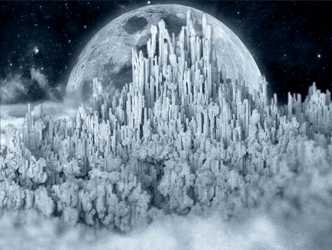
Presented vertically aligned nanostructures are Cu nanorod arrays that were prepared by template based electrodeposition method. Irregular height of the nanorods resamples rocky mountain that is lit by moonlight at mysterious night. Image width is 0.55 mm.
People’s Choice
The flower and the bud by Anurag Singhania, Gunda Harini, and Dr. Kabeer Jasuja, Indian Institute of Technology, Gandhinagar, India.
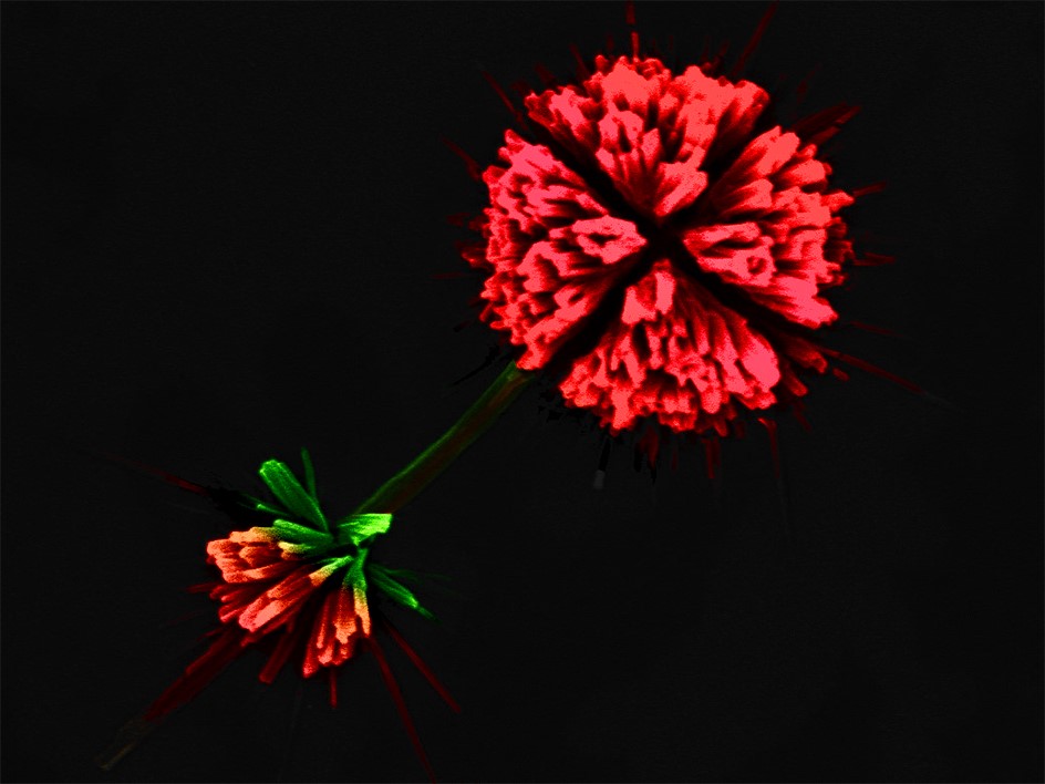
The image represents magnesium and boron based nanostructures synthesized by a bottom-up approach being developed by our research group. The phenomenon of self-assembly in the presence of specific capping agents results in formation of extremely small scale clusters which appear like growing flowers. One of the clusters resembles a full grown flower and another cluster nearby it resembles a bud in the process of growing. The clusters seem connected by a stalk like structure. This nanoflower and nanobud was imaged using a Scanning electron microscope. Image width is 0.009 mm.
Honorable Mention
Self-assembled liquid crystal flower by Wei Feng, Eindhoven University of Technology, Netherlands.

Chiral nematic liquid crystal molecules self-assemble into fingerprint pattern. In the polarized optical microscope, we can observe beautiful textures with various shapes. In the picture, there are two kinds of domains: the bright areas are planar domains and the black areas are homeotropic domains. The molecules can self-assemble into different shapes, for example, “flower-like” patterns. Image width is 0.45 mm.
Nanoyarn Meteorite by Ariana Levitt, Drexel University, USA.
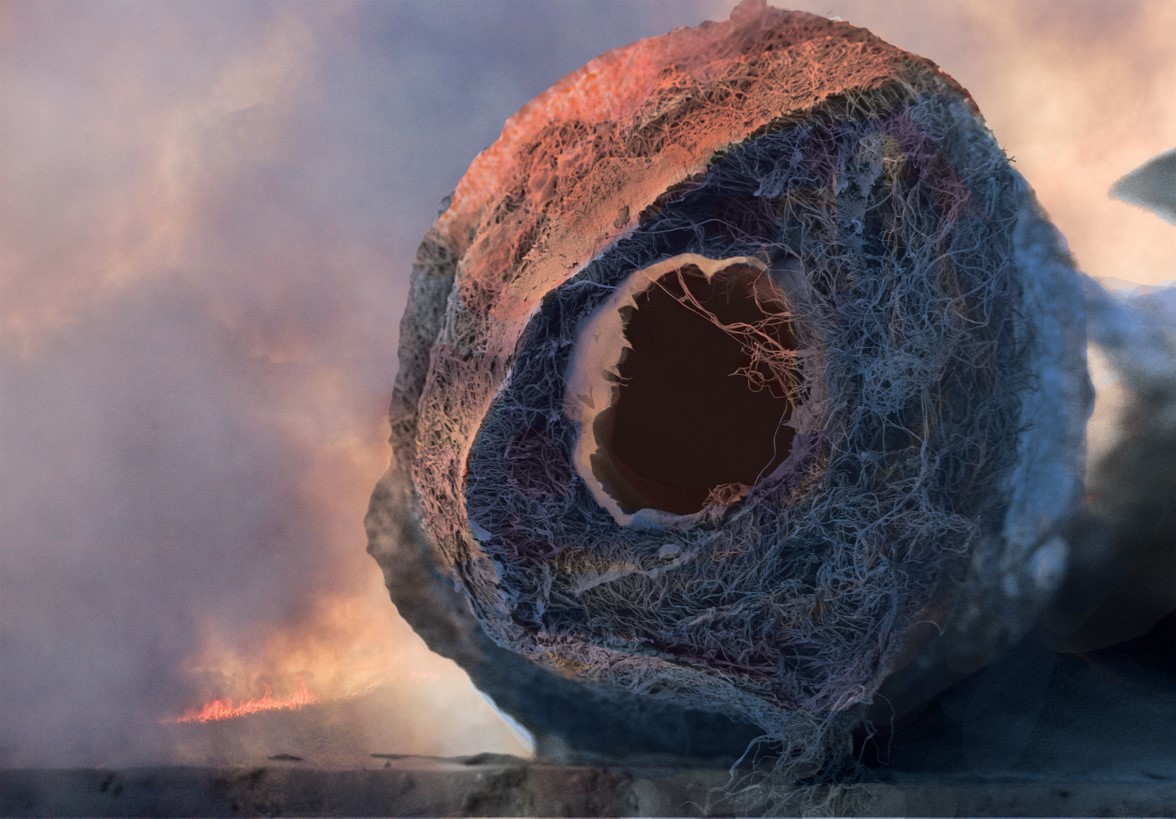
This is a false-colored SEM image of an electrospun core-shell nanoyarn, where the core is stainless steel yarn, and the sheath is composed of PAN nanofibers. PAN nanoyarns, once carbonized, are promising materials for wearable energy storage applications. Image width is 0.5 mm.
Bismuth heart by Wei Sun, Southeast University, China.
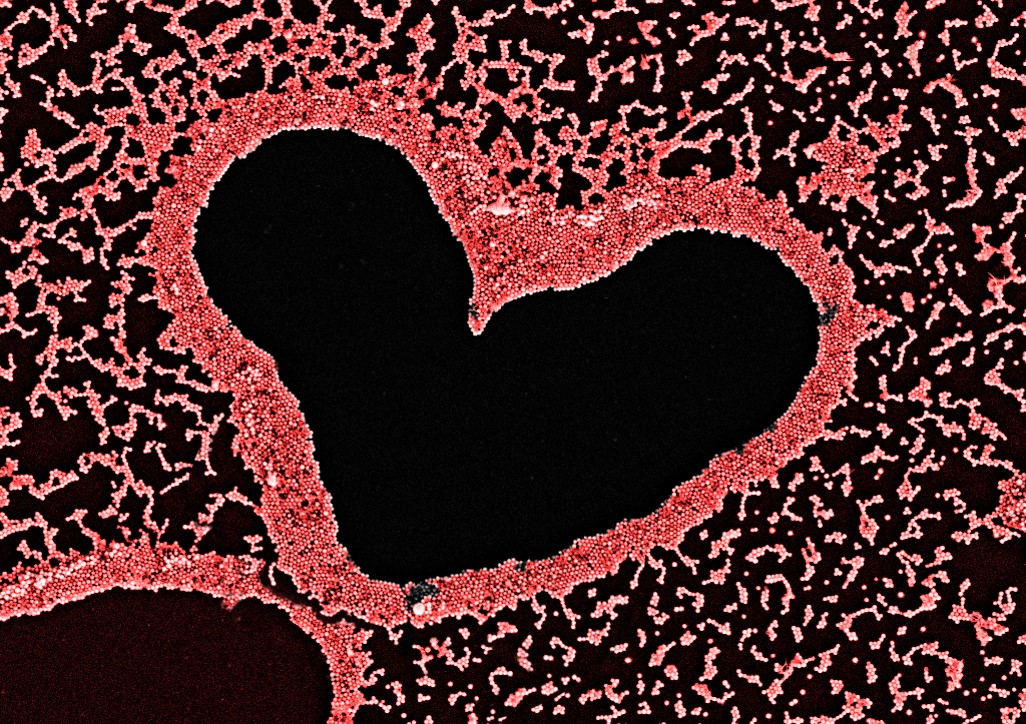
In this SEM image, we happened to find a heart-shape in a sample of bismuth nanoparticles. The special shape were formed during the drying process. Because it looks like a heart, the bismuth nanoparticles were colorized red.