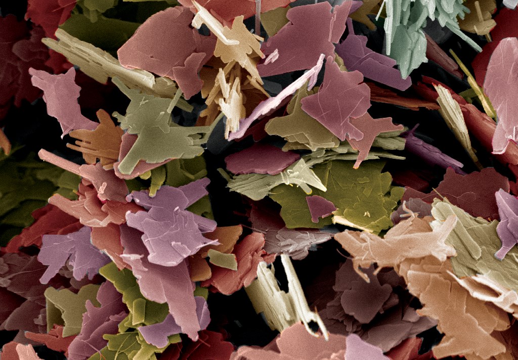
First Place
CoS2 based Nano-Micro battle stations ready for combat by Mohit Saraf, Indian Institute of Technology Indore, India.
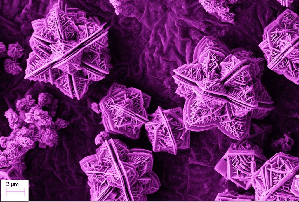
This type of never seen before morphology was grown by a simple hydrothermal reaction. Out of plethora of materials available in the literature, this unique morphology was obtained in an attempt to synthesize CoS2 nanomaterials. We believe that this type of nano-micro structure represents futuristic battle stations, which are ready to combat anytime. Scale bar is 0.002 mm.
Second Place (tie two ways)
A Polymer Whirled by Sarah Gleeson, Drexel University, Pennsylvania, USA.
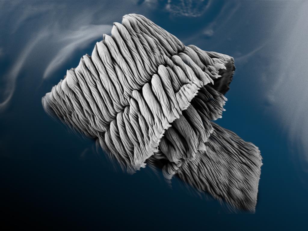
This image shows the cross-sectional edge of a polycaprolactone coating, captured with a scanning electron microscope. This coating creates a neutral surface on a polycaprolactone-block-poly (acrylic acid) single crystal film, to prevent mineral overgrowth when placed into a body-mimicking ion solution. The semicrystalline polymer film is typicallycharacterized by hard jagged faces and distinct striated lamellae. It was striking to see this portion of the coating which had coiled upon itself after being fractured, creating beautiful concentric wrinkles against a smooth polymer backdrop. Image width is 0.055 mm.
Hellfire by Jizhen Zhang, Institute for Frontier Materials, Deakin University, Geelong, Australia.
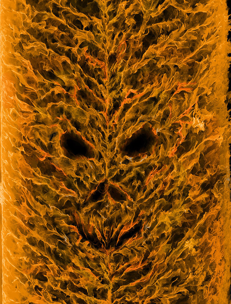
The image represents cross-section of MXene (Ti3C2Tx) aerogel film prepared by freeze-drying approach being developed by our research group. The combination of MXene flakes and porous forms flame–like architecture. The MXene aerogel film with highly aligned MXene flakes and connected interspace enable high rate charge and discharge for energy storage applications. This image was captured use a scanning electron microscope image. Image width is 0.2 mm.
Third Place (tie in three ways)
Floating Zinc Lotus by Shayan Kaviani, University of Nebraska-Lincoln, Nebraska, USA.
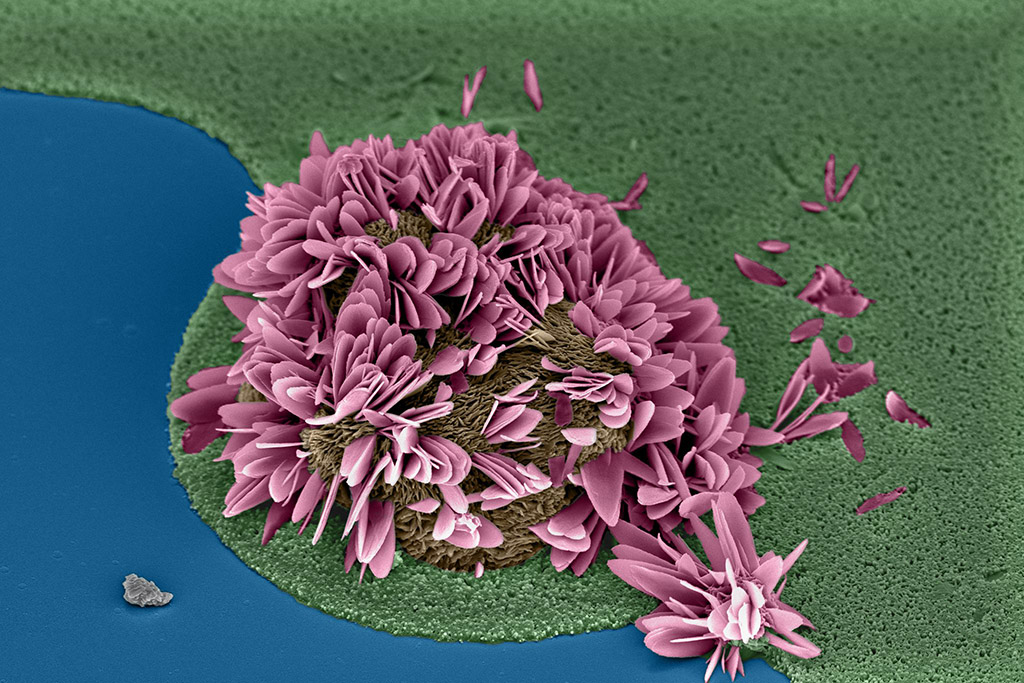
The image presents a flower-like zinc oxide nanostructure, grown on zinc-oxide nanowires, with wurtzite crystal phase. The ZnO flowers and nanowires are produced using chemical batch deposition (CBD) technique. The image is colored as a purple lotus flower floating on water. The image width is 0.030 mm.
Time for Fall: Manganese Oxide by Mallory Clites, Drexel University, Pennsylvania, USA.
Nanorods of manganese oxide are formed via hydrothermal treatment and then broken down slightly using acid leaching. The effect creates an array of small, broken fall leaves. Image width is 0.1 mm.
Nano Volcano by Simge Uzun, A.J. Drexel Nanomaterials Institute, Drexel University, Pennsylvania, USA.
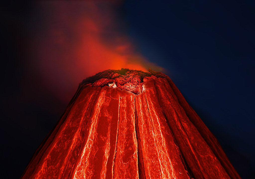
The Nano Volcano is an SEM image of a wet-spun polymer-based MXene fiber. Polyethylene glycol (PEG)
and Ti3C2 MXene dispersions are mixed in water, and the solution is used to extrude these hybrid fibers.
Here, the composite MXene fiber is represented as an active volcano, where immense amount of energy
can be released. Similarly, in this photo, we see the deeper layers of a functional fiber that has the
capability to store energy. Image width is 0.24 mm.
People’s Choice
Antman by Alberto Brambila Solórzano, Colima´s University, Colima, Mexico.
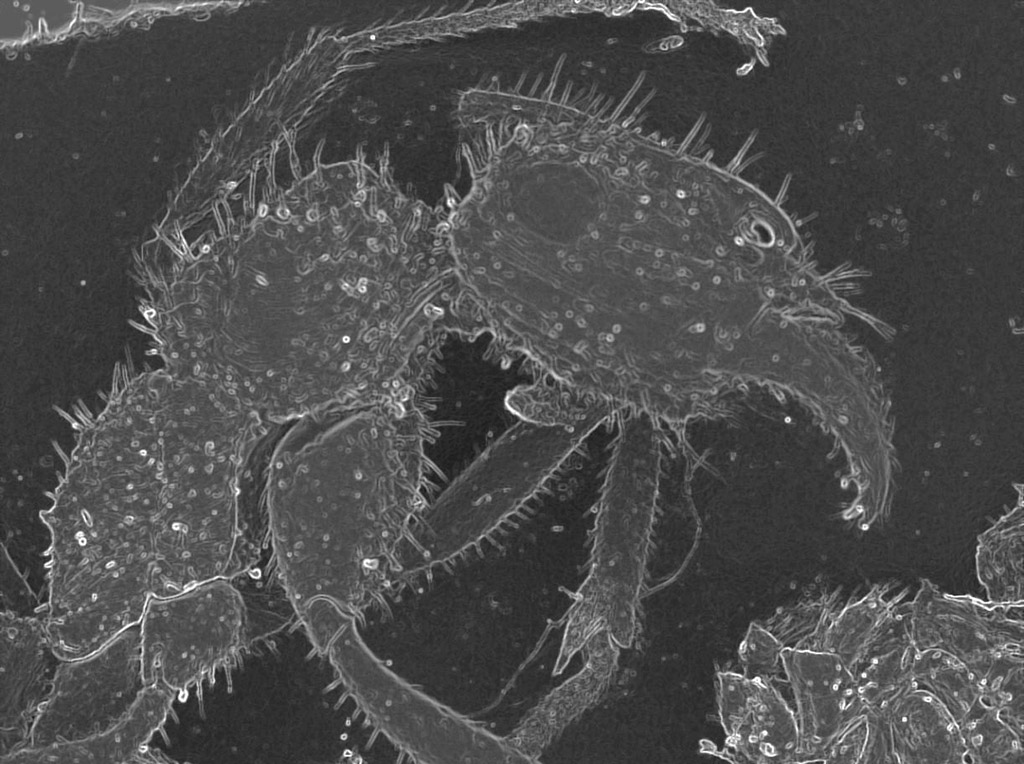
“The Antman” is a monster for his prey, in his microscopic world. It is a clear example of the beauty of nature. Width of the image is 3 mm.
Honorable Mention
A fog day in the park by Ricardo Tranquilin, CDMF/LIEC/Federal University of Sao Carlos, Sao Paulo, Brazil.
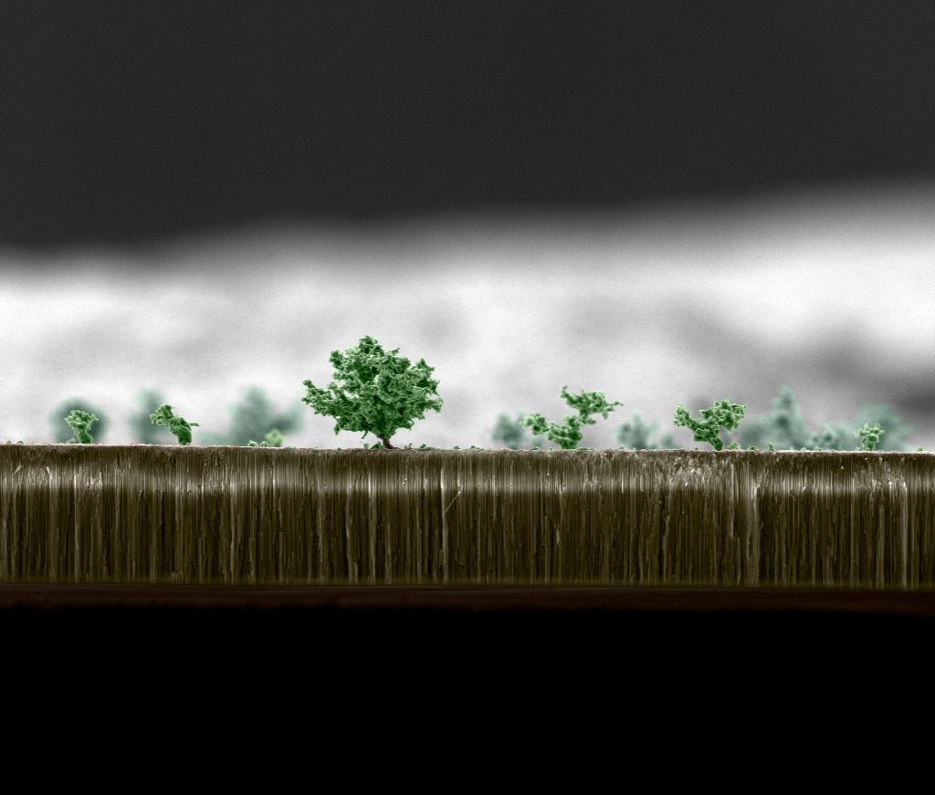
The general idea of this work is the association of nature with microscopic images, both in
their shapes and colors, so it is possible to converge the microscopic world to the comum
world, where so we can show that this small world works with the same forms of the macro
world. Also taking a special look on the images that exhibit abstract forms. The material
presented in this image is the titanium oxide deposited in aluminum oxide. Image width is 0.05 mm.
Acoustic Levitation by Aleks Labuda, Oxford Instruments, California, USA.
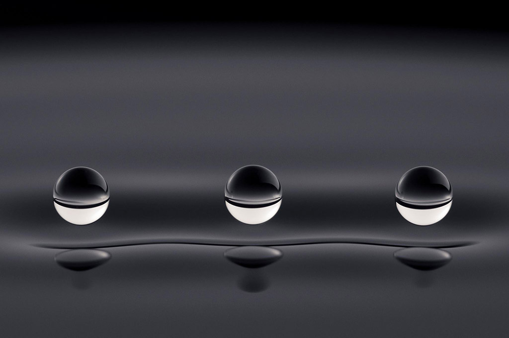
This micro-photograph depicts the first experiment in the field of hydrodynamic quantum analogs. These silicone oil droplets are bouncing indefinitely above a vibrating pool of silicone oil at 15 Hz. The surface waves generated by the droplets determine their spacing. These waves are analogous to quantum mechanical waves that guide the dynamics of quantum particles. While these droplets *move* like quantum particles, they *behave* like quantum waves. The droplets’ wave fields mediate their interactions with their surroundings, with each other, and even with themselves – similarly to electrons in the double-slit experiment.
Such bouncing droplets were discovered in 2005 by Yves Couder and mark the first real-world demonstration of the pilot-wave theory postulated by de Broglie in 1927. This behavior provides measurable and intuitive insight into the mystery of particle-wave duality. Image width is 25 mm.
MXene Lion on the Hunt by Armin VahidMohammadi and Majid Beidaghi, Auburn University, Alabama, USA.
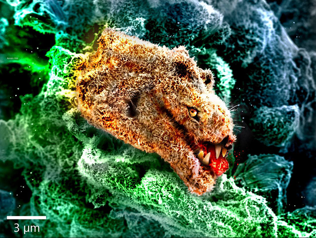
The image shows a completely oxidized two‐dimensional (2D) vanadium carbide MXene particle that resembles a head of a female lion. The image is taken with scanning electron microscopy and fake colors are added using computer graphics software. The teeth and the eye are the only things added to the image and no other manipulation has been done. Scale bar is 0.003 mm.
Drexel People’s Choice
Nano-Duck by Kanit Hantanasirisakul, Drexel University, Pennsylvania, USA.
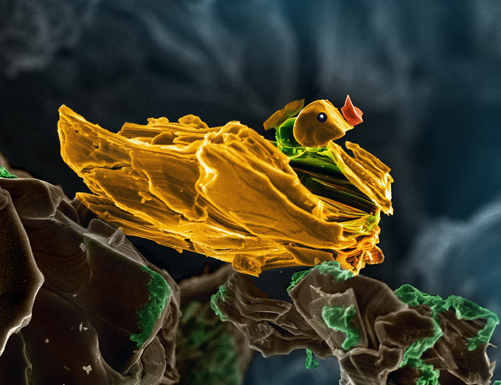
This Nano-duck is Cr2TiAlC2 MAX particle. The size of the duck is ~3 µm. The rocks where it’s sitting on are also MAX particles covered with ‘nano-moss’.