Research
Project 1: Neural circuit dynamics underlying memory consolidation
Memories are an intricate part of our life, and malfunctions in memory result in suffering such as amnesia and posttraumatic stress disorder (PTSD). Understanding how memories are formed and consolidated could provide insight into better understanding of such maladaptive memories. It is known that memories encoded during waking need to go through consolidation, a process that gradually integrates newly acquired information into preexisting neural network for long-term storage. Converging evidence suggests that the hippocampus plays a key role in this consolidation process. Damage to this structure prevents the formation of new memory, while minimally affecting old memories, indicating an interaction between the hippocampus and other brain regions for transformation of short-term memory into long-term memory.
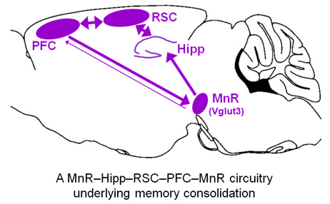
What's unclear is how exactly the hippocampus communicates with relevant cortical or subcortical regions that are also critical during the consolidation process. Of many cortical regions, the prefrontal cortex (PFC) including the anterior cingulate cortex (ACC), which is interconnected with the retrosplenial cortex (RSC), stands out as a critical site for expression of long-term episodic memory. The role of subcortical regions in memory consolidation is not well studied. We recently provided direct evidence that the mesopontine median raphe (MnR) plays a critical role in regulation of hippocampal activity and associated memory consolidation process. Based on these findings, we hypothesize that the MnR, dorsal hippocampus, RSC, and PFC form a functional circuitry during memory consolidation.
Our goal is to understand the dynamic interaction of these brain regions and the underlying neural mechanisms in the formation of a long-term memory. Results from this study will advance our understanding of memory associated circuits and provide insight for intervention into memory disorders such as amnesia and PTSD.
Project 2: Dissecting the neural circuitry of fear and anxiety en route to the amygdala
Normal fear and anxiety are important emotions within our life; however, excessive fear or pathologic anxiety can interfere with daily activities such as job performance, school work, and relationships. An estimated 40 million adults in the United States have fear- or anxiety-related disorders according to the National Institute of Mental Health, which stands as the most common of mental disorders. Thus, understanding the neural mechanisms that mediate these emotion states, fear and anxiety, is an important task in neuroscience with high relevance to human health. While fear and anxiety sometimes occur together, they are two highly distinct emotions and brain states. Fear is brief and usually evoked by a specific threat, whereas anxiety progresses and persists with no immediate threat.
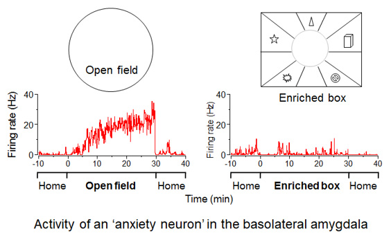
Converging evidence suggests that the amygdala is critical for processing both fear and anxiety. However, whether fear and anxiety are mediated by the same or different populations of neurons in the amygdala is unknown. Results from our recent study suggests that two groups of basolateral amygdala (BLA) neurons encode emotional states of fear and anxiety, respectively. While BLA ‘fear neurons' preferentially respond to either loud sound (auditory), moving object (visual), cat odor (olfactory), or high place exposure (multi-model) with little delay, BLA ‘anxiety neurons' gradually increase their activity under anxiogenic conditions such as exposure to a novel open field. Our goal is to determine the input and output neural circuitry of these BLA fear and anxiety neurons. A demonstration of a specific projection to BLA that regulates levels of fear or anxiety could have clinical implications for treatment of pathological fear and anxiety disorders.
Techniques
1. In vivo electrophysiology

2. In vivo calcium imaging

3. Optogenetics

4. Histology

5. Viral tracing

Back to Top