Laboratory Equipment and Instrumentation
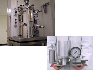 Facilities for large-scale growth of bacterial and yeast cultures include eight temperature-controlled floor shakers, a walk-in 37°C chamber, and a 20-liter New Brunswick BioFlow IV fermenter (with a dedicated CEPA continuous flow centrifuge for harvesting cell pellets). An Avestin Emulsiflex C-5 is available for rapid and efficient breakage of cells for protein purification.
Location: Department of Biochemistry & Molecular Biology
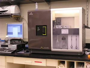 Biacore 3000 surface plasmon resonance instrument, used for detecting interactions between biological macromolecules.
Location: Chaiken Research Group, Department of Biochemistry
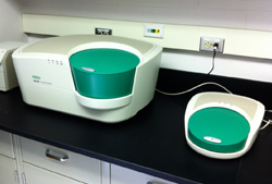 Location: Center for Advanced Microbial Processing (CAMP), 245 N. 15th Street
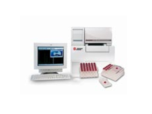 Our Beckman Coulter P/ACE MDQ capillary electrophoresis system provides a versatile, efficient, and sensitive technique for the separation of biomolecules. The option exists to pipe the output from the CE system directly to our electrospray mass spectrometer.
Location: Department of Biochemistry & Molecular Biology
Standard elevated plus maze with black walls and black runways to test anxiety.
Location: 2900 W. Queen Lane, Room 170, Barson Lab, Department of Neurobiology and Anatomy
Eight filter modes enable visible-wavelength absorption measurement applications such as protein quantification, cell viability and ELISA.
Location: 2900 W. Queen Lane, Room 170, Barson Lab, Department of Neurobiology and Anatomy
Chambers to test operant responding for liquid reinforcers.
Location: 2900 W. Queen Lane, Room 170, Barson Lab, Department of Neurobiology and Anatomy
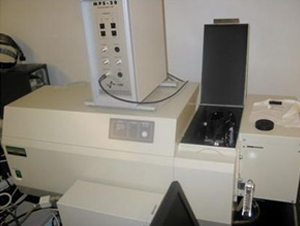 The Jorns laboratory operates a Hi-Tech Scientific Stopped-Flow System (SF-61DX2) that is equipped for anaerobic single- or double-mixing experiments with multiple detection options, including single wavelength and diode array absorbance modes.
Location: Department of Biochemistry & Molecular Biology
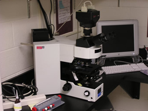 In addition, the Department has an Olympus IX71 inverted epifluorescence microscope with MetaMorph/MetaFluor Imaging System, CoolSnap HQ Digital CCD camera, and DG4 Galvo-wavelength switcher as well as an Olympus AX70 Compound epifluorescence microscope with Spot RT Slider camera.
Location: Department of Biochemistry & Molecular Biology
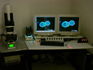 The Reginato lab operates the confocal facility featuring the Leica Laser Scanning Spectral Confocal Microscope TCS SP2 AOBS advanced image acquisition and analysis system. This system includes a UV laser, as well as standard argon, and helium neon lasers that can simultaneously image four different fluorophores. In addition, the microscope includes objectives of 20x, 40x, 63x, and 100x magnification.
Location: The Reginato Lab
 Location: Center for Advanced Microbial Processing (CAMP), 245 N. 15th Street
A high performance routine cryostat with intuitive software and touchscreen for simple, efficient operation.
Location: 2900 W. Queen Lane, Room 170, Barson Lab, Department of Neurobiology and Anatomy
Acquisition of brain activity. Calcium imaging neuroscients.
Location: Queen Lane Room 290
Acquisition of brain activity. Electrophysiologists.
Location: Queen Lane Room 290
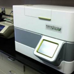 Location: Center for Advanced Microbial Processing (CAMP), 245 N. 15th Street
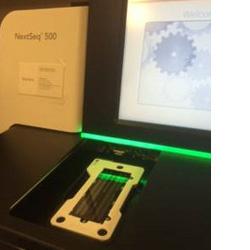 Location: Center for Advanced Microbial Processing (CAMP), 245 N. 15th Street
Fear conditioning behavioral test. Visit website to learn more. Behaviroal neuroscients.
Location: Queen Lane Room 290
The department has acquired an Odyssey infrared imaging system from LI-COR Biosciences. This is a powerful technique that can enhance the sensitivity of western blots as well as a variety of other assays.
Location: Department of Biochemistry & Molecular Biology
(w/ hole board floor inserts, two-chamber place preference inserts and light-dark box inserts) (Med Associates)
Animal movement is tracked through the use of three 16 beam IR arrays, which are located on both the X and Y axes for positional tracking and Z axis for rearing detection. Inserts allow for additional testing of novelty-seeking, reward/reinforcement and anxiety
Location: 2900 W. Queen Lane, Room 170, Barson Lab, Department of Neurobiology and Anatomy
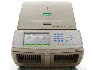 The Bio-rad CFX96™ optical reaction module converts a C1000™ thermal cycler into a powerful and precise real-time PCR detection system. This six-channel real-time PCR system comb has precise thermal control and delivers sensitive, reliable detection.
Location: Department of Biochemistry & Molecular Biology
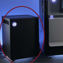 Location: Center for Advanced Microbial Processing (CAMP), 245 N. 15th Street
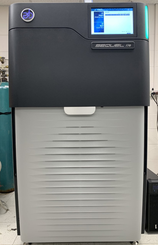
Based on the proven performance of the Sequel II system, the Sequel IIe system delivers:
- Direct access to the only highly accurate long reads: PacBio HiFi reads
- Deeper biological insights, less data processing, and faster results thanks to the unmatched clarity of HiFi reads
- Reliable and affordable high-throughput sequencing for a broad range of applications
- 8 million ZMWs per SMRTcell
Key benefits of HiFi sequencing on the Sequel IIe system:
- Better data for superior results with lower coverage, from a single technology
- Less time spent on data processing and analysis for faster answers
- Cost savings at every step of your sequencing pipeline
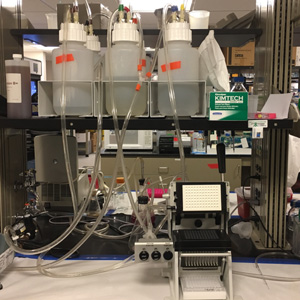 Harvests or filters cell preparations (such as transfected or stable cell lines or synaptosomes) for neurotransmitter uptake and binding assays.
Users: Pharmacology, Neuroscience and Biochemistry Students
Location: New College Building, Room 8405, Andreia Mortensen Lab, Department of Pharmacology and Physiology
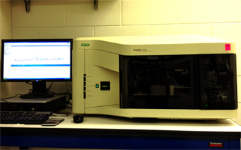 The ProteOn™ XPR36 protein interaction array system is an SPR optical biosensor that provides real-time data on the affinity, specificity, and interaction kinetics of protein interactions. Using XPR technology, a unique approach to multiplexing, this system generates a 6 x 6 interaction array for the simultaneous analysis of up to six ligands with up to six analytes. The ProteOn XPR36 system increases the throughput, flexibility, and versatility of experiment design, enabling the completion of more experiments in less time.
Location: Department of Biochemistry & Molecular Biology
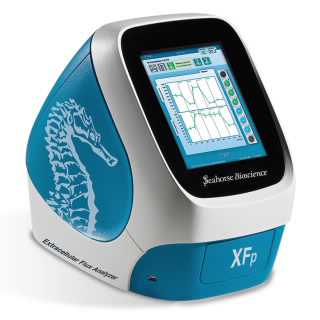 This instrument measures cellular metabolism in live cells in real time, via the measurement of extracellular flux (oxygen consumption rate and extracellular acidification rate) to provide insight into mitochondrial function and glycolytic activity. XF technology can be used to determine which pathway is predominant (metabolic phenotype), as well as which substrates are being used for energy production. Glucose oxidation, fatty acid oxidation, and aerobic glycolysis rates can be readily measured under multiple conditions.
Location: Department of Biochemistry & Molecular Biology
Full-spectrum, UV-Vis spectrophotometers used to quantify and assess purity of DNA, RNA, and protein.
Location: 2900 W. Queen Lane, Room 170, Barson Lab, Department of Neurobiology and Anatomy
A 96-well Real-Time PCR instrument utilizing robust LED based 4-color optical recording, and designed to deliver precise, quantitative Real-Time PCR results for a variety of genomic research applications.
Location: 2900 W. Queen Lane, Room 170, Barson Lab, Department of Neurobiology and Anatomy
Simultaneously disrupts multiple biological samples through high-speed shaking in plastic tubes with stainless steel, tungsten carbide, or glass beads. Using the appropriate adapter set, up to 48 or 192 samples can be processed at the same time. Alternatively, a grinding jar set can be used to process large samples.
Location: 2900 W. Queen Lane, Room 170, Barson Lab, Department of Neurobiology and Anatomy
Back to Top