The Sanz-Clemente Lab
The Sanz-Clemente Lab is devoted to identifying molecular mechanisms regulating glutamate receptor subsynaptic location and utilizing this knowledge to revert/prevent alterations associated with synaptopathies. To achieve this goal, we utilize a combination of in vitro and in vivo approaches, including biochemistry, pharmacology, genetics, cellular/molecular biology, state-of-the-art imaging techniques, and –omics approaches. Our current interests include:
Molecular mechanisms controlling the balance between synaptic and extrasynaptic NMDARs
NMDA Receptors (NMDARs) are a kind of glutamate receptors that mediate the calcium-permeable component of excitatory neurotransmission. Post-synaptic NMDARs are segregated in two pools: synaptic (sNMDARs) and extrasynaptic (exNMDARs). Although both s- and exNMDARs are stable populations and display distinct, independent functions, they are connected via regulated lateral diffusion. Physiologically, this exchange contributes to “NMDAR-plasticity”: the bidirectional modulation of the synaptic NMDAR content in an activity-dependent manner. This process controls metaplasticity, homeostatic plasticity, and some modulatory functions, such as the integration of synaptic input. Despite its importance, the mechanisms that govern the regulated s/exNMDAR exchange are poorly understood. Based on previous findings, we hypothesize that s- and exNMDARs display a distinct post-translational modification (PTM)-profile that, in combination with a specific set of interacting proteins, determine NMDAR subsynaptic localization. Specifically, we are studying a particular phosphorylation on the GluN2B subunit of NMDARs (on S1480) and the interaction of GluN2B with SAP102 and Kif5b as key determinants for controlling s/exNMDAR balance.

A model for s/exNMDAR balance regulation: Activation of sNMDARs (1) activates calcium-calmodulin kinase II (CaMKII) and promotes CaMKII interaction with GluN2B. (2) Activated CaMKII also binds to casein kinase 2 (CK2) (3) to facilitate the CK2-mediated phosphorylation of the GluN2B PDZ-ligand (at S1480, GluN2B-pS1480) (4). GluN2B-pS1480 disrupts NMDAR binding to MAGUKs (4) allowing their lateral diffusion to extrasynaptic sites (red arrow). The clearance is dependent on the interaction of GluN2B with SAP102 in a secondary (non-PDZ) binding site (5). ExNMDARs are internalized and, subsequently, recycled (black arrow). However, a portion of exNMDARs become stabilized at the extracellular cell surface (red box) in a GluN2B-pS1480-manner (6). GluN2B-pS1480-containing exNMDARs form a protein complex including inactive protein phosphatase 1 (PP1) (7). Co-activation of s- and exNMDARs (1)+(8) drives PP1 activation (9) and dephosphorylation of GluN2B-pS1480 (10). Dephosphorylated NMDARs are reintegrated to the postsynaptic density via lateral diffusion (green arrow) where they become stabilized via PDZ interaction with MAGUKs.
Inverse correlation between the levels of GluN2B-pS1480 and GluN2B-containing NMDAR synaptic content: Levels of GluN2B-pS1480 were pharmacologically modulated in acute hippocampal slices from >P70 mice. The reduction in the levels of GluN2B-pS1480 (blue dots) is associated with an elevated presence of GluN2B-containing NMDARs at biochemically isolated synaptic sites. Conversely, induction of GluN2B-pS1480 (red dots) promotes GluN2B synaptic clearance, resulting in a decreased GluN2B synaptic content.
Modulation of synaptic NMDAR content as a novel therapeutic strategy to reduce excitotoxicity
Excitotoxicity is defined as the deterioration of the neuronal function/structure caused by excessive glutamatergic stimulation. It is a converging pathomechanism observed in many neuropathies, including Alzheimer’s disease (AD), Huntington’s disease (HD), amyotrophic lateral sclerosis, and traumatic brain injury (TBI), among others. The overactivation of exNMDARs is required for excitotoxicity as the dysregulated Ca2+ entry into the synapse via exNMDARs impairs mitochondrial function, activates calpain, and inactivates survival signaling. Therefore, exNMDARs are obvious pharmacological targets to prevent/reduce excitotoxicity. Unfortunately, inhibiting exNMDARs without affecting sNMDARs is challenging, and apart from memantine, NMDAR antagonists have failed in clinic due largely to the side effects caused by sNMDARs inhibition. We are currently evaluating a novel therapeutic strategy based on the modulation of the s/exNMDAR balance rather than on the inhibition of NMDAR activity. We hypothesize that modulating NMDAR lateral diffusion to recruit exNMDAR to synaptic sites and/or prevent NMDAR synaptic clearance would reduce excitotoxicity by (i) reducing pro-death signaling (exNMDAR-mediated) and (ii) promoting survival cascades (sNMDAR-mediated). We are initially testing this innovative strategy based on reducing excitotoxicity by modulating NMDAR trafficking in animal models of AD.
Chronic inhibition of casein kinase 2 (CK2) in primary neuronal cultures alters the transcriptome profile associated with cell survival. Primary neuronal cultures were treated with the selective CK2 inhibitor CX-4945 for 3 weeks, and their transcriptome profile was analyzed using Illumina HiSeq 4000. RNA-seq analysis reveals a profound transcriptomic reorganization that includes several signaling pathways associated with neuronal survival.
Biochemical isolation of synaptic plasma membranes (SPMs) from primary neuronal cultures (+/- NMDAR activation) reveals a stable pool of protein phosphatase 1 (PP1) associated with the plasma membrane.
Investigating the molecular mechanisms underlying synapse unsilencing during development and its influence in Rett syndrome pathophysiology
GluN2B-containing NMDA receptors regulate important developmental processes in the central nervous system such as synaptogenesis, synaptic activation, synaptic maturation, and dendritic arborization. Accordingly, GRIN2B, the gene encoding GluN2B, is one of the few genes scored with “high significance” to be associated with autism spectrum disorder (ASD) risk. Despite the solid genetic evidence, how GluN2B function/trafficking is affected in ASD remains unknown. We hypothesize that alterations in the post-translational modification (PTM)-profile of GluN2B at early developmental stages contribute to ASD pathogenesis by altering GuN2B-containing NMDAR trafficking. Our preliminary data have identified dysregulated levels of a specific phosphorylation on GluN2B (on S1323) in animal models of Rett syndrome, the leading cause of severe intellectual disability in girls. We found that, physiologically, GluN2B S1323 phosphorylation regulates GluN2B surface expression and controls synapse activation and dendritic morphology, two of the points of convergence for ASD. We are currently investigating the potential amelioration of pathological outcomes observed in animal models of Rett syndrome by normalizing the levels of S1323 phosphorylation.
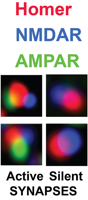
Identification of active (AMPAR-containing) and silent (AMPAR-lacking) synapses utilizing structured illumination microscopy (SIM) super-resolution microscopy.
Back to Top