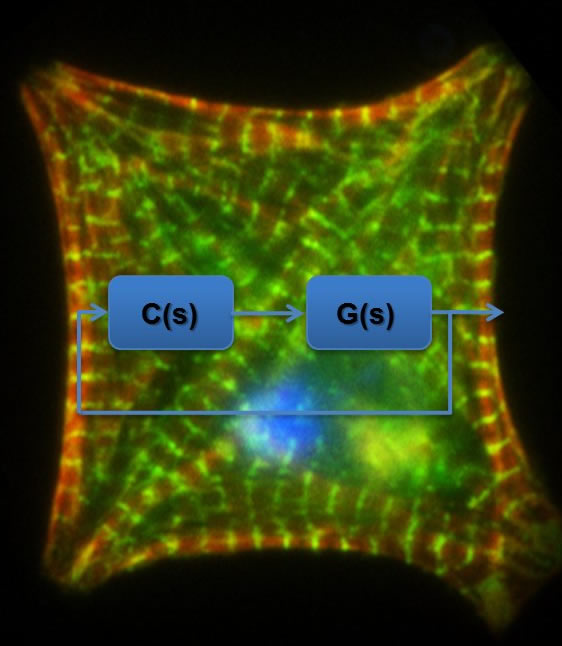
Cell-matrix and cell-cell adhesions are crucial to the maintanance of the structural integrity and contractile function of cardiac myocytes. Changes or disruptions to these adhesions can have adverse affects on myocyte remodelling (shape and cytoskeletal architecture) resulting in loss in mechanical and electrical syncytium as seen in heart failure. Studies in mechanobiology have focused on attachment of sub confluent cells to ECM ligands. It has not been determined whether N-Cadherin (N-Cad) acts as a mechanosensor. To test the hypothesis that N-Cad is directly involved in mechanotransduction, neonatal ventricular myocytes are plated on a model gel system emulating cells of varying stiffnesses, functionalized with N-Cad and ECM. Cells are interrogated with atomic force microscopy (AFM) and labelled for sarcomeric Α -actinin and F-actin, vinculin and beta catenin. Our early results indicate that cells in contact with soft (300 Pa) N-Cad gels, myocytes do not develop F-actin fibers and are devoid of sarcomeric organization. At physiological meaningful tissue stiffness (15 kPa), cells display striated F-actin and organized myofibrils while in contact with stiff (scar-like) gels (60 kPa), cells display prominent F-actin filaments without striations. These results show, for the first time, that changes in N-Cad mediated traction forces can alter the cytoskeletal organization in a manner similar to integrins. Importantly, these results have broad implications in understanding remodeling associated with heart failure and therapies such as mechanically unload of the heart (e.g. ventricular assist devices, constrainment).