Olympus FV1000 - Laser Scanning Confocal
The Cell Imaging Center houses two Olympus FV1000. One is located in PISB Room #408 and the other Bossone Room #625C.
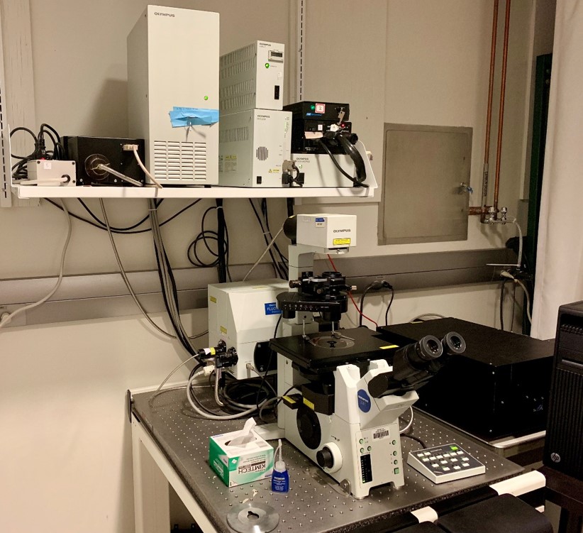 The Olympus FV1000 is an inverted microscope - objectives are below the stage. This confocal microscope is ideal for 2D or 3D visualization and quantification of specimens ranging from single cells and tissues to small organisms (e.g., Drosophila larvae, C. elegans).
The FV1000 is capable of simultaneous and sequential high-resolution imaging of three or more fluorophores.
Differential Interference Contrast (DIC) is also available to acquire a contrast image of a transparent sample.
The FV1000 system supports state-of-the-art imaging applications including colocalization, volume rendering, particle tracking and motion analysis, fluorescence recovery after photobleaching (FRAP), fluorescence loss in photobleaching (FLIP), and fluorescence resonance energy transfer (FRET).
Light Sources
Halogen lamp for bright-field microscopy (white light illumination)
- a contrast image of the specimen can be directly observed through the eyepiece
Mercury arc lamp for direct visualization of fluorescent specimens through the eyepiece
Laser lines
- 405 nm for DAPI staining
- 458 nm for CFP
- 488 nm for GFP / FITC / Alexa 488 / Cy2
- 515 nm for YFP
- 543 nm for tdTomato / mCherry / TRITC / Cy3
- 635 nm for Alexa 647 / Cy5
Objectives
(Magnification / Numerical aperture)
- 10x/0.4 Dry (Olympus)
- 20x/0.75 Dry (Olympus)
- 40x/0.85 Dry (Olympus)
- 40x/1.3 Oil (Olympus)
- 60x/1.42 Oil (Olympus)
- 100x/1.4 Oil (Olympus)
Detectors
3 Hamamatsu photomultiplier R7862 tubes (PMTs).
Acquisition software
FV10-ASW version 02.01 (Olympus)