The Drive to Find a Solution

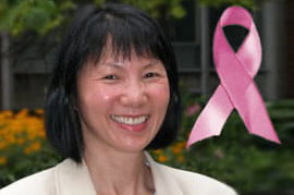
In honor of Breast Cancer Awareness month, BIOMED associate professor Dr. Wan Shih shares with DrexelNow her personal story of breast cancer survival, which was the inspiration behind her research to develop technology to more quickly and effectively detect breast cancer in women.
In 2008, Dr. Wan Shih was too busy for a biopsy. After a routine mammogram, her doctor saw something suspicious, ordered follow-up, and then recommended a biopsy. But Dr. Shih had too much going on.
“Then, a colleague of mine said if I didn’t go and get the biopsy, he would no longer work with me,” she explains.
That colleague is known as the friend who saved her life.
Three years later, Dr. Shih feels lucky to say she’s cancer free. She wants that outcome for all breast cancer sufferers, and knows early detection is crucial. The problem is that mammography has some flaws.
Dr. Shih explains that mammography is not always effective with women who have dense breasts, a common physiological feature among Asian women and a large percentage of women in the U.S. aged 40 to 50. She and her team at Drexel developed a non-invasive, radiation-free, portable device used for breast cancer detection based on measurements of tissue elasticity. The device is inexpensive, easy to use and sensitive enough to provide clues as to whether a lump is benign or malignant, potentially eliminating some of the many biopsies performed every year.
A hand-held probe comprising piezoelectric finger (PEF) sensors can detect very small forces and displacements at the surface of the breast, she adds. These are then converted to electrical signals that can help detect even very small tumors, and also differentiate cancers from non-cancerous lumps.
In addition to its effectiveness as a physical detection device, rather than an imaging tool like mammography, the device’s portability is important in developing countries where mammography is not widely available.
Early studies have shown that, in several cases, Dr. Shih’s device detected cancerous tissue when mammography did not.
“We scanned 40 women and 11 of them had cancerous-type pathology. Our device detected all of them and mammography missed some,” she says. “This proof, of course, is still preliminary, but it’s encouraging that this device can pick up cancers that are missed by mammography.”
Dr. Shih says her latest research deals with breast-cancer imaging during surgery, a technique that would help reduce the number of patients requiring second surgeries. The imaging takes place and is analyzed after the first surgery, and if any cancerous tissue remains, it is removed, thereby eliminating the need for a second surgery in most cases.
Dr. Shih knows there is much to be done before getting her screening device to market. But the motivation to work hard and meet the challenge is strong and constant.
“I feel if I can do something meaningful with the rest of my life, then I will be happy,” she says.
Dr. Shih developed the breast cancer screening device with Drs. Wei-Heng Shih (BIOMED) and Ari Brooks (DUCoM), who will be presenting the technology at the American College of Surgeons Clinical Congress Oct. 23-27. The device was licensed by UE LifeSciences, a Philadelphia-based medical device company focused on bringing effective and sustainable breast cancer solutions to life, and the project received funding through the University City Science Center’s QED Program in 2009. The project also received support from the Wallace H. Coulter Translational Research Program at Drexel.
Read more here.
Drexel News is produced by
University Marketing and Communications.