Gross Anatomy: Now and Then
- by guest blogger, Chrissie Perella, iSchool grad student and intern extraordinaire
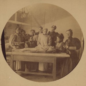
Woman's Med students in the gross anatomy lab, 1897
Several days ago, we were fortunate to get a tour of the gross anatomy lab here at Drexel University College of Medicine. And by gross, I don't mean disgusting - I mean what the medical world terms macroscopic; that is, things that can be identified and measured with the naked eye.
Our tour guide was a gracious and knowledgeable first-year med student. The students are divided into groups of 5 or 6 and each group is assigned their own cadaver. The only information they receive on the body is the sex, age, profession, and cause of death. During the course of the fall and spring terms, the students carefully dissect their cadavers, all the while learning about the delicate nature of the human body - organs are removed, muscles examined, and arteries and veins followed. Students must be careful to keep their cadavers preserved by covering them with moist towels and a bag following every class. Our guide said she felt very fortunate to be able to learn in such a hands-on way. A little known (but nice) fact: An event honoring the donors and their families is held at the end of year. Read about the event on philly.com: "Students pay tribute to body donors: Honoring the ultimate gift," Philadelphia Inquirer, 28 April 2010
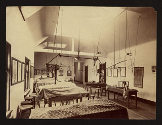
The cadaver lab of Woman's Medical College of Pennsylvania, 1895.
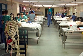
The cadaver lab at College of Medicine, 2011. Photo courtesy of the Drexel Med Emergency Medicine Residency Program
And all this left me wondering: what was dissection like for earlier students at the Woman's Medical College? So, here's a look back at Woman's Med students in the gross anatomy (cadaver) lab:
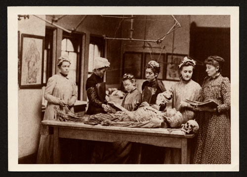
Comparing anatomical drawings to real life, circa 1892.
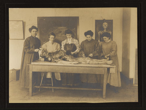
No gloves needed for dissection in the early 1900s! (circa 1901)
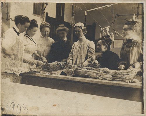
A lesson being taught in the cadaver lab, 1903.
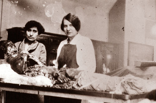
Two medical students working hard at learning anatomy, 1928.