Total Internal Reflection Fluorescence Microscopy
Principle
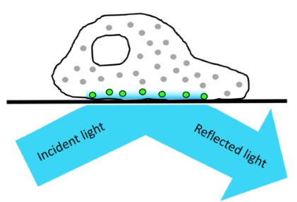
 The DeltaVision OMX V4 enables Total Internal Reflection Fluorescence Microscopy (TIRF). This technique uses an evanescent wave to selectively excite fluorophores that are close to the coverslip. This evanescent wave is generated when the incident light is totally reflected at the interface between the glass coverslip and the specimen. The intensity of the evanescent electromagnetic field falls off exponentially from the interface. As a result, the excitation of fluorophores is restricted to a region that is ~100 nm deep from the coverslip.
Advantages
The main advantage of using TIRF is its outstanding signal-to-noise ratio due to the low penetration depth of the evanescent field. Therefore, out-of-focus fluorescence is dramatically minimized and almost no background fluorescence occurs. In addition, this restricted excitation greatly reduces photobleaching, which improves cell viability.
TIRF is a wide-field technique so images can be rapidly acquired on a camera chip, and multiple fluorophores can be imaged simultaneously or sequentially.
A drawback of TIRF microscopy is the necessity to use adherent cultured cells; tissue slices, for example, are generally not close enough to the coverslip/medium interface.

Applications
TIRF has been used to study the spatio-temporal dynamics of many different molecular processes at or near the cell membrane. Therefore, TIRF has become the method of choice for imaging processes occurring at the bottom of live cells such as cell adhesion, endo-exocytosis, and cytoskeletal dynamics.
In vitro TIRF assays are also widely utilized to study the interaction of diffusible ligands with biomolecules immobilized on functionalized glass substrates.