Super-resolution
Principle
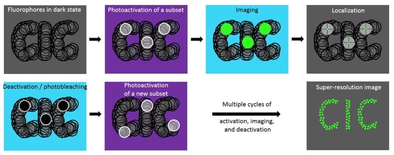
The DeltaVision OMX V4 enables Localization Microscopy (LM), a method that breaks the diffraction limit of light. In conventional microscopy, fluorophores are simultaneously excited in a sample, making it difficult to spatially resolve closely spaced fluorophores. In contrast, LM resolves individual fluorophores temporally. LM takes advantage of fluorescent dyes or proteins that undergo light-induced photoactivation or photoswitching. By turning on and off each individual fluorophores, LM is able to identify their position with a high degree of precision. Once the centroid of each fluorophore has been determined, a final image is reconstructed with nanometer precision.
Advantages
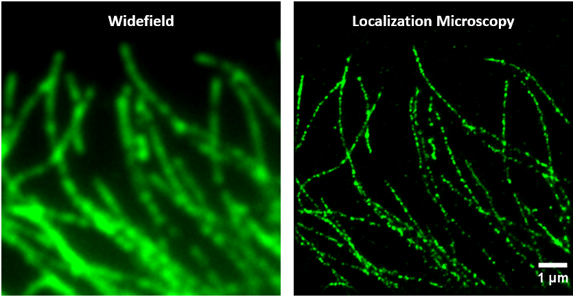
LM is the super-resolution technique that achieves the best spatial resolution. LM improves the lateral resolution by an order of magnitude over conventional microscopy (20-50 nm vs 250 nm).
Limitations
Because only a small subset is turned on at a time, thousands of images must be acquired to reconstitute the final image of the specimen. Several minutes are often necessary to acquire a super-resolution image, which limits its use to fixed specimens.
In addition to this low temporal resolution, LM is limited by its practical imaging depth (~200 nm in TIRF mode), and its reliance on a computationally derived image that may introduce artifacts. In addition, multicolor imaging is challenging.
Location
Drexel University
Papadakis Integrated Sciences Building
4th Floor
3245 Chestnut Street
Philadelphia, PA 19104