Wide-field
The Cell Imaging Center houses two wide-field microscopes: the Olympus IX81 and the Zeiss AxioObserver.
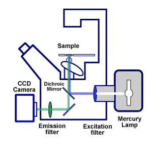
A wide-field microscope is a type of fluorescence microscope in which the entire specimen is illuminated by a white light source, usually a mercury arc lamp. Optical filters are used to generate excitation light of specific wavelengths. Excitation light is directed to the sample via a dichroic mirror, which reflects some wavelengths but transmits others. The emitted fluorescence can be viewed directly through the eyepiece or projected onto an image capture device, which is usually a Charge-Coupled Device (CCD) camera.

In wide-field microscopy, secondary fluorescence emitted by the specimen from above and below the focal plane interferes with the resolution of the features in focus. As a result, high-magnification wide-field images of specimens having a thickness greater than ~2 µm appear blurry. Thus, wide-field fluorescence microscopy is not optimal for 3D imaging. However, out-of-focus haze can be removed computationally after acquisition by applying deconvolution algorithms to 3D stacks of images.
