Cell Imaging Center
Mission
Our mission is to provide a state-of-the-art facility, expertise, and education in advanced quantitative light microscopy to support Drexel University’s research community
The CIC
The Cell Imaging Center (CIC) is a multi-user facility housed in purpose-built imaging suites in the Papadakis Integrated Sciences Building (PISB) and in the Bossone Research Center. The CIC serves the light microscopy needs of Drexel researchers from a variety of academic colleges and departments including the College of Arts and Sciences, the College of Engineering, the School of Biomedical Engineering, and the College of Medicine. The CIC also supports the teaching missions of Biology and Biomedical Engineering curricula and provides service to users from local biotech companies.
Vision
Inspire innovative research and empower Drexel’s researchers through acquisition and promotion of emerging advanced imaging technologies, excellent service, and community partnerships
Values
Inclusion | Excellence | Integrity | Innovation | Access | Stewardship | Education
Services
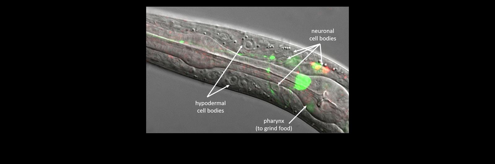
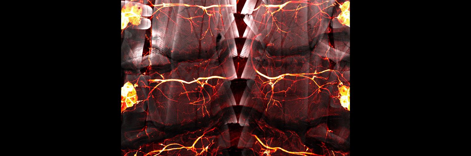
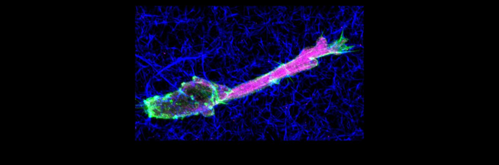
The CIC provides researchers with state-of-the-art light microscopes to visualize and quantify a variety of cellular and molecular processes within fixed or living cells and tissues. This is accomplished using a variety of optical imaging techniques including:
- Wide Field microscopy / Stereology
- Laser Scanning Confocal microscopy
- Two-Photon Excitation microscopy
- Super-resolution microscopy (3D Structured Illumination Microscopy and Localization Microscopy)
- Total Internal Reflection Fluorescence microscopy (TIRF)
- Fluorescence-Activated Cell Sorting (FACS)
Location
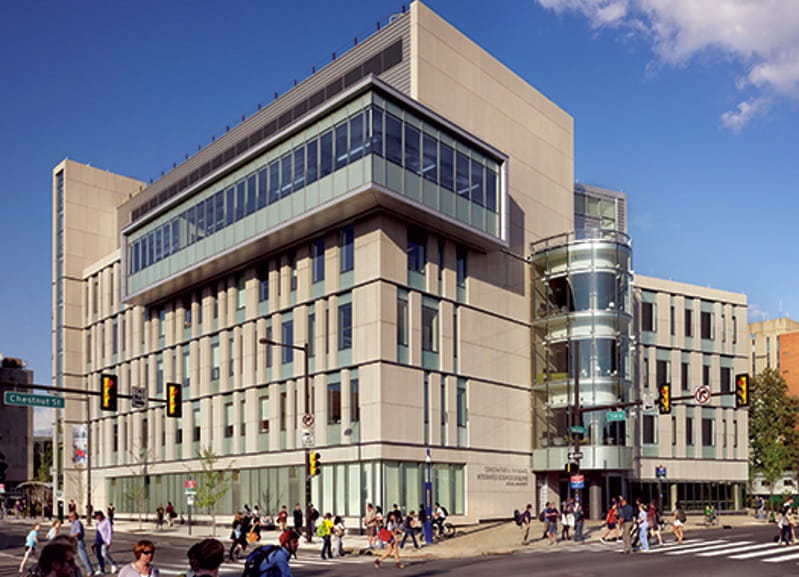
Drexel University
Papadakis Integrated Sciences Building
4th Floor
3245 Chestnut Street
Philadelphia, PA 19104