DeltaVision OMX V4 – Multi-mode Super-Resolution Microscope

The DeltaVision OMX V4 from Applied Precision (GE Healthcare) is a fully automated inverted microscope - objectives are below the stage. The OMX is a multi-mode imaging platform that enables imaging of fixed or live specimens at better spatial resolution than conventional microscopy (wide-field and confocal microscopy).
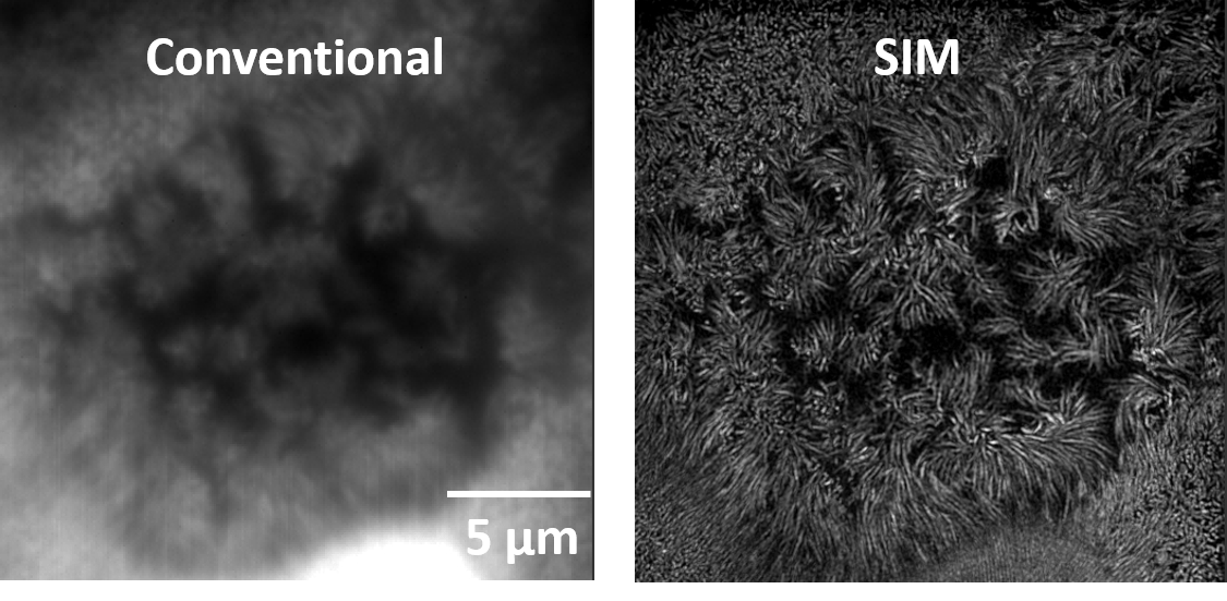
Due to the wave nature of light, any point source of light (even from a single GFP molecule with a size of 3 nm) is observed by the microscope as a small diffracted blur. As a result, the spatial resolution (i.e. ability to distinguish 2 points) is limited to ~250 nm in the x-y plane and ~600 nm in the z-direction. Compared to conventional microscopy, the OMX super-resolution microscope improves spatial resolution by a factor of 2 to 5.
The OMX is equipped with a structured illumination microscopy (SIM) module, which enables 3D super-resolution imaging of live and fixed samples at ~120 nm lateral and ~300 nm axial resolution using standard fluorophores.
The OMX also contains a DeltaVision localization microscopy (DLM) module, which achieves imaging of fixed samples at a 20-50 nm lateral and 100-150 nm axial resolution using photoactivatable and photoswitchable fluorophores.
The OMX imaging platform also offers TIRF (Total Internal Reflection Fluorescence) microscopy, which illuminates fluorophores within ~100 nm depth from the coverslip-sample interface without interference from out-of-focus light.
For live cell imaging, a “Ultimate Focus” module maintains samples at the focal plane over time, and a temperature-controlled chamber and objective warmer maintain optimal environmental conditions.
A better spacial resolution, but what for?
Super-resolution imaging techniques have become commercially available in the past 5 years and their use will continue to grow exponentially. Super-resolution is revolutionizing many different biological and biomedical research fields. The reason why is simple: we can now visualize and study molecular assemblies and structures that were not accessible to conventional light microscopes.
Examples of applications include the study of micro-organisms (e.g. bacteria, viruses), organelles (nucleus, mitochondria, vesicles), protrusions (filopodia, spines, cilia), plasma membrane organization (lipid rafts), and structural organization of large protein complexes (cytoskeleton, synaptic protein, nuclear pore complex).
Light Sources
Halogen lamp for bright-field microscopy (i.e. white light illumination to acquire a contrast image)
Solid state illumination module for wide-field fluorescence microscopy capable of simultaneous operation of 4 user-selectable wavelengths from 6 possible excitation wavelengths (405, 445, 488, 514, 568, and 642nm)
Laser lines for TIRF, 3D SIM, and Localization microscopy:
- 405 nm for DAPI staining
- 445 nm for CFP / Alexa 430
- 488 nm for GFP / FITC / Alexa 488 / Cy2
- 514 nm for YFP
- 568 nm for tdTomato / mCherry / TRITC / Alexa 568 / Cy3
- 642 nm for Alexa 647 / Cy5
Objectives
(Magnification / Numerical aperture)
- 60x/1.42 (Olympus)
- 60x/1.49 (Olympus) – for TIRF
Detector
3 pco.edge sCMOS cameras (custom version for DeltaVision OMX V4)
Acquisition software
OMX Master (GE)
Image reconstruction and analysis software
softWoRx 6.1.1 (GE)
Location

Drexel University
Papadakis Integrated Sciences Building
4th Floor
3245 Chestnut Street
Philadelphia, PA 19104