To the extent that all scientific research relies on technology, the next great discovery may be at the other side of a latest-generation lens. For researchers at Drexel University College of Medicine, the acquisition of two new confocal microscopes provides an unprecedented view into the human body — and fresh insights into conditions such as autism, neuroinflammation, HIV-associated neurocognitive impairment, heart attacks, cancer and more.
Confocal microscopy deploys laser scanning for optical imaging. Through multiple scans of sections of the object with a controlled depth of focus, the microscope and its associated software can assemble and digitally render a three-dimensional view. While the College has owned confocal microscopes for many years, the newest versions, purchased at the end of 2016, offer updated features such as higher resolution, better accuracy, low-temperature operability and greater speed for processing data. A Leica microscope has already been installed in the Department of Neurobiology & Anatomy at the Queen Lane Campus while an Olympus model is going into the New College Building on the Center City Campus, with both being made available to multiple investigators.
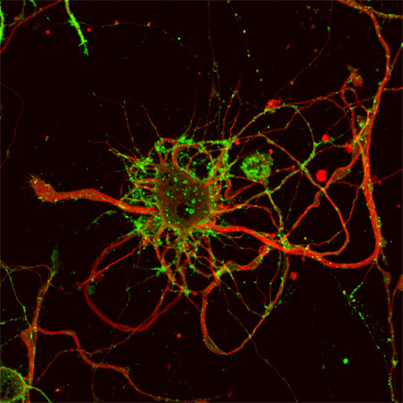
Confocal image of a cortical neuron expressing the herpesvirus transport protein US9 fused to a green fluorescent protein, which allows the visualization of specific membrane microdomains (i.e., lipid rafts).
The investment in the equipment allows for a more efficient and powerful approach to research. "In the past it would take us up to two hours to analyze a single neuron, and now we can do that at least three times faster," says Olimpia Meucci, MD, PhD, chair of the Department of Pharmacology & Physiology. The improved pace not only speeds up the course of an investigation, but it also makes it possible for more investigators to access the equipment on a given day and for greater spans of time. Meucci estimates that double the number of investigators can now use the equipment to look at live cells.
The level of detail in the new scopes' imaging enables new understanding of cell processes at the molecular level. "In my lab, we look at changes in the intracellular localization of specific proteins involved in neuroprotection, as well as their interaction with other proteins and cellular structures," Meucci says. "One of my students has just started a series of experiments dissecting the molecular pathways implicated in the regulation of dendritic spines — tiny protrusions of the membrane receiving inputs from other neurons — in cortical neurons. Another student is looking at the effect of HIV viral proteins on neurons and how we can exploit the herpes simplex protein US9 as an intracellular carrier to deliver ‘therapeutic tools' into affected neurons. Both of these projects require long-term study of live neurons and a complex imaging analysis that would not be feasible without the new confocal," she says.
There is great excitement and enthusiasm in the Department of Pharmacology & Physiology for the new scope's capabilities, says Harpreet Singh, PhD, an assistant professor. "We can now go macro to micro with high sensitivity and better temperature controls. The Olympus can also perform super resolution, which is a bonus," says Singh. In his lab, investigators are looking at single mitochondria from the heart, isolating these 1-to-2-micrometer organelles in order to study the role they play in fine-tuning the physiological impact under pathological conditions such as heart attacks.
The newer models allow investigators to examine live cells with greater stability. In the Department of Neurobiology & Anatomy, Assistant Professor Kazuhito Toyooka, PhD, is studying brain development in transgenic mice and, specifically, how neurons in the cerebral cortex and hippocampus migrate. "I watch in vivo cells in time-lapse imaging over 24-hour periods to analyze their gene function during brain development. In the past, I would have to mark the cells with a strong signal, the laser would bleach out the image, and it was challenging to keep the samples stable. Now, with the high-sensitivity detectors in the new microscope, we can see every detail and observe the samples for longer stretches of time." One result, he hopes, will be an understanding of the genes linked to conditions such as autism and schizophrenia and how they develop in the brain.
One of the most exciting possibilities for the new instruments is that they may pave the way for greater cross-department collaboration, Meucci says. "It's not simply an update for our facilities. The new acquisitions show the College's commitment to attracting the highest quality students and faculty, increasing exchange between basic science and clinical medicine for greater research outcomes."
The advances in equipment and their new capabilities will also spur wholly new paths of inquiry. "Just a few years ago it was not even feasible to imagine looking at mitochondria on plates without looking at the whole cell," Singh says. "Now it's what we're doing every day. This technology has enabled me to create my own niche to study the ion channels present in organelles. Seeing is believing."