
Background
Hometown: Richmond, Virginia
Undergraduate: Virginia Commonwealth University, BS in Clinical Laboratory Sciences
Graduate: Drexel University College of Medicine, MS in Medical Science
Q&A
Can you tell me a little bit about yourself as far as your background before you came to Drexel?
I'm originally from Virginia, and I went to undergrad at Virginia Commonwealth University. I graduated with a degree in clinical laboratory sciences. For about seven years, I worked for the VCU Health System as a medical laboratory scientist in their chemistry lab, running blood tests and fixing their chemistry analyzers.
I came to Drexel's master's in medical science program looking to make a career change from laboratory science into medicine. I wanted to use this program as a way to leverage myself, improve my application, and improve my chances of being accepted into medical school.
What was attractive about Drexel's program?
I really liked that it was in an urban city. I actually applied to the Drexel Pathway to Medical School program but didn't get accepted. However, they shifted my application materials over to the Medical Science program and I was accepted. It actually worked out great because this program really fits my needs.
Are you in your second year now?
Yes. I'm in my second year. This is actually my last semester, so I'm really excited. I've had one medical school interview so far at Marshall University in West Virginia. Hopefully, that'll work out and I'll get accepted, but I'm still focused on my research here and really excited about it. I'm doing research on septins. It's exciting because it's pretty cutting edge. If some of my experiments work out, we could really have something that could be worthy of being published in a journal.
Can you tell me more about your research?
Septins are a cytoskeletal protein system that are the most recently discovered and least studied. There's about 13 septin genes. You have individual or septin monomers. They assemble into complexes of either six or eight polypeptides. The one with six polypeptides is known as a septin hexamer. The one with eight polypeptides is the octamer. These complexes then assemble end to end to make septin filaments. That's the crux of what I'm studying.
The octamer contains Sept9 and the hexamer does not. Sept9 is overexpressed in various cancers such as colorectal and breast cancer, to the extent that methylation of the SEPT9 gene is an FDA approved circulating biomarker for colorectal cancer screening. The mechanism by which the elevated Sept9 protein levels contribute to these cancers is not understood making my research of septin dynamics imperative.
My research experiment will start the process of unraveling septin dynamics by clearly delineating whether the septin octamer can co-polymerize with the septin hexamer. To accomplish this, I am purifying and fluorescently labeling different septin assemblies from bacteria, and exploring their polymerization using fluorescence microscopy.
This experiment will guide many others by defining if Septins should be thought of as a single filament network or as multiple distinct filament networks within the cell.
How do you conduct your research?
We insert septin genes into bacteria using DNA recombinant tools than express the septin proteins in E. coli. Once we've put our plasmid into E. coli, we grow them, they express our protein, and then, after they express it, we purify the protein. Once we've purified it, the hope was to label it, but we actually found a way to label it inside the DNA gene itself so that when it's expressed, it already has this part that we added onto it that is a fluorophore. We don't have to label it because it has its own synthetic label. The fluorophores we're using are GFP and mCherry.
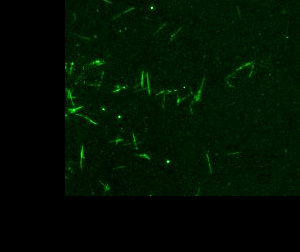
Once we've expressed it and it's got the fluorophore on it, we're going to use confocal microscopy, or this other microscopic technique known as TIRF, to image it. I'm hoping next week to have some really neat, nice images of one filament that's going to be labeled red and one that's going to be labeled green. If they're two separate systems, when I look on the microscope, I'll see red filaments and green filaments, and they won't mix. They'll be distinct. However, if they are one system, they'll share monomers, so when I look under the microscope, I'll see yellow or orange filaments because they've been interchanging.
Do you have any advice for someone who is considering doing the medical science program here?
Understand the curriculum and the goals of the program so that you know if it's the right fit for you, because it's definitely not for everyone. It is a rigorous program, but I definitely feel like I've gained a lot of tools. I feel very comfortable doing research. I feel very comfortable presenting it and creating PowerPoints, so I've definitely gained a lot from the program. It's definitely worthwhile. Just make sure you're ready for the rigor, and go for it. It's a good program.