Gene Therapy Research
We use recombinant retrovirus and adenovirus vectors to deliver neurotrophin genes as a strategy of promoting survival of injured neurons and axon regeneration. The recombinant retroviruses and adenoviruses contain a neurotrophin transgene and a reporter gene, which allows their travel to be tracked in tissue. The virus vectors are injected into the spinal cord and migrate both rostrally and caudally from the site of injection. As the virus migrates from the injection site, a gradient of neurotrophin gene expression is produced. Our theory is that this gradient will encourage injured host cells to project axons towards the site of injection and grafted cells to project axons away from the site of injury. We have since used similar strategies to deliver neurotrophins and chondroitinase (to digest CSPG) with lentiviruses and AAV.
Injection of recombinant adenovirus into spinal cord
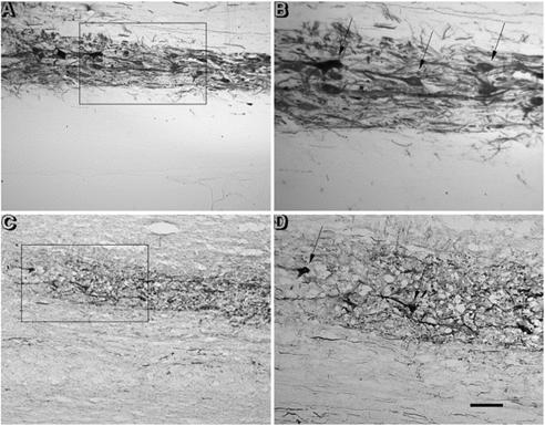
Fig. 1 Transgene expression at the injection site, one week following surgery. The injection site shows intense staining for lacZ transgene expression. A,B: X-gal histochemical staining. C, D: immunocytochemical staining obtained with an anti-β-galactosidase (β-gal) antibody and DAB as the chromagen. Staining is localized to the injected side with little staining on the contralateral side. At higher magnification (B and D), both staining methods demonstrate detailed morphology of cell bodies and processes, resembling a Golgi staining patters (arrows). Scale bars: A, C = 100μm; B,D = 40μm.
Transgene expression in distal regions after 1 week and 2 months
Fig. 2. Transgene expression distant from the injection site. Transgene expression was detected by X-gal histochemistry (A-H) or β-gal immunocytochemistry (I). One week following injection, expression level is high, resulting in Golgi-like staining patterns in neurons of the red nucleus (A and B), locus coeruleus (D and E), reticular formation (G) and propriospinal neurons (H). The staining is mostly localized to the ipsilateral locus coeruleus and Clarke's nucleus and the contralateral red nucleus. In A, the crossing rubrospinal fibers are clearly visible (arrows). Two months following injection, expression level is greatly reduced and staining is localized only to cell bodies, as shown for red nucleus (C), locus coeruleus (F), and Clarke's nucleus (I). Scale bars: A, D = 250μmm; B, C, E, F, H = 100μmm; G = 40μm; I = 125 μm.
Related Publication
Application of recombinant adenovirus for in vivo gene delivery to spinal cord. [PDF]
Liu Y1, Himes BT, Moul J, Huang W, Chow SY, Tessler A, Fischer I.
Brain Res. 1997 Sep 12;768(1-2):19-29.
Back to Top