One small misstep in the biomechanical understanding of the ankle about 70 years ago has thrown research on the joint slightly off kilter ever since. Sorin Siegler, PhD, a mechanical engineer at Drexel is hoping to realign thinking surrounding one of the busiest orthopedic intersections in the body.It all started about three years ago when Siegler was attempting to make a cast of the outside of the talus bone – the pivot bone of the three that make up the ankle.
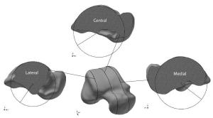
“We did a number of CT scans of the ankle, slicing it in to small sections and then used a computer program to make a three-dimensional model of the talus,” Siegler said. “The project was to replicate the outer shape of the bone so that it could be used in ankle replacements.”
What Siegler found was that the slices of the inner part of the ankle –the medial side- were larger in diameter than the slices on the outer –lateral- side. While the significance of the observation didn’t immediately strike him, when he looked at the 3-D models it became increasingly obvious that something was off.
“When we laid out all the 3D CAD renderings, we simply placed them on the page without a particular orientation, aside from aligning it vertically to position medial and lateral sides appropriately,” Siegler said. “Looking at the images this way made it easy to see that the talus formed a truncated cone with its apex on the lateral side.”
This all may seem like a pedestrian, academic study of orthopedics – of some importance to people who study ankles or who injured one, but not to the rest of the walking world. What difference does it make that a 3D computer rendering of a bone, not more than few inches big –that appears, with a bit of imagination, to look like a cone with its top lopped off- actually points toward the outside part of the leg instead of the inside? Quite a bit. A finding like Siegler’s actually cuts to the core of the scientific method.
From Observation to Fact
To understand why this discovery is so significant, you have to take a look at the work of Verne Thompson Inman, who literally wrote the
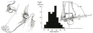
book on ankles, and originally published this work in 1952. Inman postulated a fixed axis of rotation for the ankle and he based his measurements of the talus, conducted by hand from actual slices of the bone recovered from a cadaver, on this premise.
And this is how he went about constructing the idea that the talus bone has an overall conical shape and the apex of that cone is on the inside of the foot, the medial side. Over the course of 70 years of orthopedic and biomechanical research this theory became an immutable –or at least unquestioned- fact about the ankle.
Retracing Steps
During this time period, scientists have come to new understandings about how the ankle moves and, to a degree, what it looks like. But the one element that never changed in 70 years was the idea of the conical talus with its apex on the medial side. The problem with this assumption, as Siegler discovered, is that Inman’s measurements were based on the ankle maintaining a fixed-axis orientation.
One thing that modern ankle research, including Siegler’s, has shown, is that the ankle axis is far from fixed. Siegler’s modeling has demonstrated that the coronal plane of the talar dome –the top of the ankle bone that connects with the leg- has a saddle-like indentation. This explains how the leg-to-ankle connection allows the foot to rotate on a flat plane, as well as pronating and supinating –that is, landing on the outside or inside part of the foot when walking.
With this in mind, if Inman’s measurements are redone without assuming the fixed axis of ankle rotation-which was essentially what Siegler’s model did- the apex of the cone clearly appears to be on the lateral side –the exact opposite of Inman’s finding.
A Realignment
So what does this all mean? It doesn’t change the way people have walked for thousands of years, so what’s the big deal?
“What this does, is change the way we understand and attempt to recreate the movement of the ankle,” Siegler said. “Artificial ankle replacement operations have been performed for less than a decade, there is still plenty of room for improving the technology and this could be a big part of that improvement.”
The next steps for this research could help people with degenerative ankle problems, like arthritis, take their next steps. Having a better understanding for the shape and movement of the bones in the ankle will allow for a more accurate fabrication of joint replacements and, as a result, a more natural walking motion for people who use them.
Siegler’s team is in the process of making an ankle implant with surface geometry that follows his hypothesis. In the coming months they will put a 3D-printed replicate of a state-of-the-art prosthesis up against a Siegler-designed prototype in a series of biomechanical tests –that include implanting both prosthetics in a cadaver ankle to see which produces a more natural ankle movement.
But Siegler contends that the greatest significance of this finding –no matter what it holds for the future prosthetics- is that it reminds us to be constantly inquisitive.
“This process has taught me a lesson that all scientists should, but not always follow. In science, as perhaps in other areas in life, you must question and probe everything!” Siegler said. “Sometimes the most established theories, opinions and views of our world can turn out to be wrong when probed clearly against reality using the tools available to us."